For decades, the “Big Three” of cancer treatment were surgery, chemotherapy, and radiation. Today, we have a fourth pillar and that is Immunotherapy. Instead of using chemicals to attack the tumor directly, immunotherapy empowers your own immune system to fight back and kill.
What is Immunotherapy?
Immunotherapy is a biological therapy. Our immune system is already designed to detect and destroy “foreign” invaders like bacteria and viruses. However, cancer cells are masters of disguise—they can “hide” from the immune system or send signals that switch off the bodies defence.
Immunotherapy can help treatment in three ways that is,
- Marking cancer cells, so they are easily found by immune system.
- Boosting the immune system to work harder against the disease.
- Eliminating the “brakes” used by cancer cells to stop immune
There are several ways doctors “train” the immune system to fight back. Here are the most common methods:
1. Immune Checkpoint Inhibitors
The immune system detects “checkpoints” to prevent attacking healthy or self cells. Cancer cells often hijack these checkpoints to stay safe. Checkpoint inhibitor drugs block these checkpoints, giving the immune system the “green light” to attack.
- Example: Pembrolizumab and Nivolumab. These are widely used for advanced melanoma and non-small cell lung cancer.
To understand Immune Checkpoint Inhibitors (ICIs), you have to think of the immune system not just as a weapon, but as a vehicle. For the vehicle to move safely, it needs both an ignition switch (to start an attack) and brakes (to stop it from attacking its own body).
Cancer cells are essentially “hijackers” that slam on the body’s molecular brakes, forcing the immune system to stand down while the tumor grows. ICIs work by cutting those brake lines.
a. The Biological “Brakes”: How Checkpoints Work
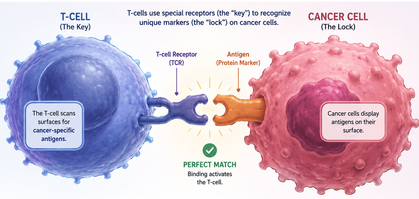
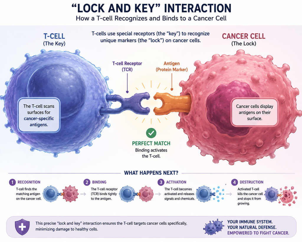
In a healthy body, T-cells (the “soldiers” of the immune system) use checkpoint proteins to determine if a cell is a threat or a friend.
When a T-cell’s receptor binds to a healthy cell, these checkpoints act as an “off switch.” This prevents autoimmunity—where your body accidentally attacks itself. The two most common “brakes” are:
- CTLA-4 (Cytotoxic T-Lymphocyte Associated Protein 4): Acts early in the process, usually within the lymph nodes, to prevent T-cells from being “turned on” in the first place.
- PD-1 (Programmed Cell Death Protein 1): Acts later, typically at the site of the tumor, to stop T-cells from actually killing the cells they find.
b. The Cancer “Hijack”: PD-L1
Cancer cells evolve to produce a protein called PD-L1. When a T-cell approaches a tumor, the PD-L1 on the cancer cell binds to the PD-1 on the T-cell.
- The Result: The T-cell receives a signal that says, “I’m a normal cell, don’t hurt me.” , ultimately the T-cell becomes “exhausted” or inactive, and the tumor continues to grow undetected.
c. The Mechanism of the Inhibitors (The Drugs)
Immune Checkpoint Inhibitors (ICI) are monoclonal antibodies (lab-made proteins). They work through a “lock and key” mechanism:
- Blocking the Interaction: The drug molecules physically sit in the “lock” (the PD-1 or CTLA-4 receptor).
- Neutralizing the Signal: Because the drug is occupying the receptor, the cancer cell’s PD-L1 can no longer bind to it.
- Reactivation: The “off” signal is never sent. The T-cell remains active, recognizes the cancer cell as a threat, and begins its attack by releasing toxic granules to destroy the tumor.
d. Why Isn’t It Perfect?
Since, ICIs “remove the brakes” from the immune system, the T-cells can sometimes get a bit over-enthusiastic. If they stop recognizing healthy tissue as “self,” they can cause Immune-Related Adverse Events (irAEs). This essentially looks like temporary autoimmune diseases, such as:
- Colitis (inflammation of the colon)
- Pneumonitis (inflammation of the lungs)
- Dermatitis (severe skin rashes)
The beauty of this mechanism is its durability. Because it teaches the immune system what the cancer looks like, some patients experience “memory” responses where the cancer stays away for years after treatment ends.
2. CAR T-Cell Therapy
CAR stands for Chimeric Antigen Receptor.
- Chimeric: Because the receptor is made of parts from different sources (like the Chimera of mythology).
- Antigen: The “target” on the cancer cell.
- Receptor: The “hook” that grabs the target.
Doctors remove a patient’s T-cells (a type of white blood cell), genetically engineer them in a lab to make them recognize a specific protein on cancer cells, and then infuse them back into the patient.
- Example: Tisagenlecleucel, used primarily for certain types of leukemia and lymphoma.
While Checkpoint Inhibitors (the ones we discussed earlier ) focus on “unbreaking” the immune system, CAR T-cell Therapy is more like building a custom-made “super-soldier” to hunt down a specific enemy.
It is often called a “living drug” because the treatment is made of your own living cells, which continue to grow and multiply inside your body.
The Manufacturing of CAR-T cells
The process is incredibly complex and personalized for every single patient:
- Apheresis (Collection): Doctors draw blood from the patient, then separate out the T-cells (the immune system’s primary attackers). The rest of the blood is returned to the patient.
- Reprogramming (The Lab): Scientists use a deactivated virus to insert a new genetic blueprint into the T-cells. This blueprint instructs the cells to grow “CARs” on their surface.
- Expansion (The Greenhouse): Those few engineered T-cells are grown in the lab until they grow into hundreds of millions.
- Conditioning: Before the cells are put back, the patient usually undergoes a mild round of chemotherapy to clear out some existing immune cells, making “room” for the new super-soldiers.
- Infusion (The Attack): The CAR T-cells are dripped back into the patient’s bloodstream.
How It Works Inside the Body
Once the CAR T-cells are back in the body, they act as guided missiles:
They recognise, binds and kills the cancer cells, and itself proliferates.
i. Recognition
Normal T-cells need help from other cells to “see” cancer. CAR T-cells don’t. Their new receptors act like a GPS, specifically tuned to find a protein on the cancer cell (most commonly one called CD19 found in B-cell leukemias).
ii. Binding and Killing
When the CAR “hook” finds the cancer “antigen,” it locks on tightly. This binding sends a massive signal to the T-cell to activate. The T-cell then releases toxic chemicals to punch holes in the cancer cell, killing it instantly.
iii. Proliferation
The best part? When a CAR T-cell hits its target, it starts cloning itself. One T-cell can turn into thousands, creating a massive army that scours the body for every last trace of cancer.
The “Cytokine Storm”
Because CAR T-cells are so powerful, they can cause a side effect called Cytokine Release Syndrome (CRS). As the cells rapidly kill cancer, they release a flood of chemicals (cytokines) into the blood. This can cause high fevers and low blood pressure—it’s actually a sign that the treatment is working, but it requires careful hospital monitoring.
THIS THERAPY IS MOSTLY USED OR BLOOD CANCER, AND NOT FOR SOLID TUMORS.
- The Problem: Solid tumors are dense, physical masses. They are surrounded by a “fortress” of connective tissue called the stroma.
- The Result: CAR T-cells often struggle to physically penetrate the tumor. They stay on the outside, unable to reach the cancer cells at the core.
3. Monoclonal Antibodies
These are synthetic proteins designed to bind to specific target on cancer cells. Some “flag” the cancer so the immune system can see it, while others block signals that tell the cancer to grow.
- Example: Trastuzumab (Herceptin), which targets the HER2 protein in some breast cancers.
Monoclonal antibodies (mAbs) like Trastuzumab (Herceptin) are precision-engineered proteins. While CAR T-cell therapy is like a “soldier” and Checkpoint Inhibitors are like “cutting the brakes,” Trastuzumab is more like a highly specific “clamp” that locks onto a target to disable it.
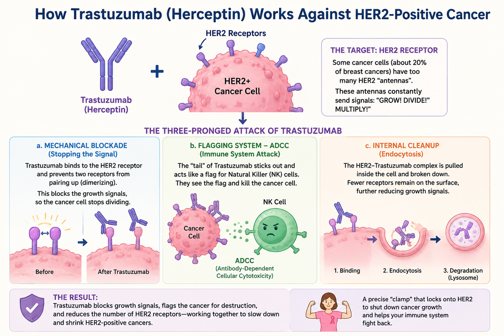
How monoclonal antibodies work against HER2-positive cancers.
i. The Target: The HER2 Receptor
Some cancer cells, especially in about 20% of breast cancers have an abnormally high number of HER2 (Human Epidermal Growth Factor Receptor 2) proteins on their surface.
Think of these HER2 receptors as antennas. In a healthy cell, they tell the cell when to grow. In cancer, there are too many antennas, and they are constantly screaming “GROW! DIVIDE! MULTIPLY!”
ii. The Three-Pronged Attack of Trastuzumab
Trastuzumab attacks the cancer in three distinct ways:
a. The “Mechanical Blockade” (Stopping the Signal)
Normally, HER2 receptors need to pair up or dimerize to send a growth. Trastuzumab physically attaches itself to the outside part of the HER2 receptor and acts like a wedge, preventing the receptors dimerization. This “muffles” the growth signals, and the cancer cells stop dividing.
b. The “Flagging” System-Antibody-Dependent Cellular Cytotoxicity (ADCC)
This is an immunotherapy like activity of the drug. Once Trastuzumab is clamped onto the cancer cell, its “tail” sticks out into the bloodstream. This tail acts as a giant neon sign for Natural Killer (NK) cells. Immune system sees the “flag” and realizes this cell is a target. This process is called Antibody-Dependent Cellular Cytotoxicity (ADCC)— In simpler words the drug points the finger, and the immune system pulls the trigger.
c. Internal Cleanup (Endocytosis)
When the drug binds to the receptor, it sometimes triggers the cell to pull the receptor inside itself to be broken down. There are fewer “antennas” left on the surface to receive growth signals, further weakening the tumor.
2026 Context: Antibody-Drug Conjugates (ADCs)
By 2026, we’ve moved beyond just “plain” Trastuzumab. We now frequently use ADCs like Trastuzumab Deruxtecan (Enhertu).
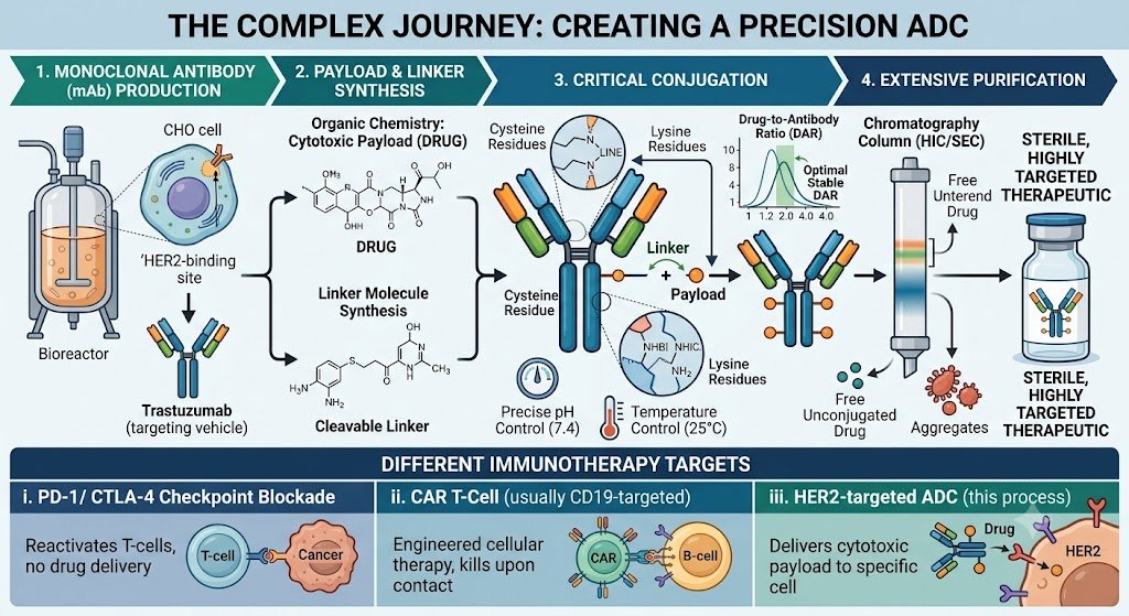
Antibody-drug conjugates (ADCs) are made of complex multi-step process that begins with the production of a monoclonal antibody (mAb), which serves as the targeting vehicle. Simultaneously, the cytotoxic payload (DRUG) and the chemical linker are synthesized through specialized organic chemistry. The critical “conjugation” step follows, where the linker and payload are chemically bonded to specific sites on the antibody—such as cysteine or lysine residues—using precise pH and temperature controls to ensure a stable Drug-to-Antibody Ratio (DAR). Finally, the mixture undergoes extensive purification, often via chromatography to remove unconjugated “free” drugs and any unstable aggregates, resulting in a sterile, highly targeted therapeutic.
4. Cancer Vaccines
Unlike flu vaccines that prevent disease, most cancer vaccines are therapeutic—they help the immune system attack an existing tumor. Example: Sipuleucel-T (Provenge), used for some men with advanced prostate cancer. For cancer vaccine information and possibility of its success, stay tuned, Another blog will soon be available.